
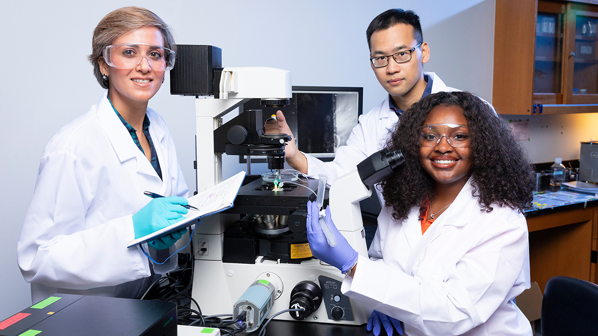
(Left-right): Postdoctoral researcher Rojan Saghian, Ph.D., assistant professor in chemical & biomedical engineering Z. Leonardo Liu, Ph.D., and undergradtuate student Leah DuBose pose with a microscope in a laboratory at FAMU-FSU College of Engineering in Tallahassee, Florida. The team plans to create a biophysics-informed AI framework that infers blood properties from videos of blood flow and uses them to assess disease risk. (Scott Holstein/FAMU-FSU College of Engineering)
Key Points
Z. Leonardo Liu, an assistant professor of chemical and biomedical engineering at the FAMU-FSU College of Engineering in Tallahassee, Florida, is leading a new National Science Foundation project to measure how blood flows through the body’s smallest vessels.
The team will combine microfluidic chips, computer simulations and physics-informed artificial intelligence to infer blood properties such as thickness, cell stiffness and clotting tendency from videos of a small blood sample.
Current methods require a large blood draw and several specialized lab instruments. The researchers aim to develop a fast, affordable test that could help doctors track conditions such as sickle cell disease and clotting disorders at the bedside.
Standard blood tests can diagnose sickle cell disease, but they can’t predict when a patient’s cells will jam a tiny vessel. A FAMU-FSU engineer is training AI to read that risk from a single drop of blood.
Abnormal blood clotting and other blood disorders contribute to heart attacks, strokes and life-threatening complications for millions each year. Physicians can analyze many aspects of blood, but still cannot routinely measure how blood behaves in the body’s tiniest blood vessels. Known as the body’s microcirculation, this is where many diseases begin and take hold.
Researchers at Florida A&M University and the FAMU-FSU College of Engineering are launching a study to change that with a new National Science Foundation project, “AI-Powered Data Framework for Probing Microscale Blood Flow.”
The project is led by Z. Leonardo Liu, an assistant professor of chemical and biomedical engineering at the FAMU-FSU College of Engineering. Liu currently has a postdoctoral fellow and an undergraduate student on his team.
Their approach combines microfluidic chip experiments, computer simulations and physics-informed artificial intelligence to probe the mysteries of microvascular blood flow. “With just one drop of blood, we can learn the underlying characteristics that drive complications of conditions such as sickle cell disease and clotting disorders,” Liu said.
“Standard tests can diagnose sickle cell disease, but they can’t predict how likely a patient’s red blood cells are to get stuck in tiny vessels on a given day,” he added. “By analyzing blood flow at a microscopic level, we hope to learn not only a patient’s condition, but also their risk of complications, such as a blocked vessel, in the days ahead.”
How does blood behave in the body’s smallest vessels?
Blood behaves differently in the body’s tiniest vessels. “Many of blood’s most important jobs happen in tiny vessels so narrow that red blood cells must squeeze through one after another, and that’s where many diseases start,” Liu said.
At the microscopic scale, blood is nothing like a simple liquid such as water. It is a crowded mixture of flexible cells that squeeze through narrow vessels, rearrange and sometimes stick together.
“If we can observe how blood moves at that microscopic scale, we can work backward to what is actually driving the disease—whether the cells are too stiff, too sticky or prone to getting jammed up,” Liu said.
Why can’t current blood tests measure microscale blood flow?
In traditional imaging, scientists can watch blood in motion. However, they can’t see what the blood is like “rheologically,” meaning how thick it is, how stiff or sticky the cells are, or how easily clots can form.
Currently, obtaining this information requires a sizable blood draw and several specialized lab instruments. These include a rheometer, the benchmark tool for measuring blood viscosity, along with separate tests for cell stiffness and clotting.
How will AI measure blood properties from a single drop?
Liu’s team is taking a different approach. “Instead of relying on a large blood draw and bulk lab instruments, we flow a small sample through tiny channels on a microfluidic chip, record videos of how it moves, and use AI to infer the blood’s properties from that motion,” Liu said. The result, he added, is “a quick, simple measurement that tells you what the blood is like at the cellular level.”
The team plans to build a biophysics-informed AI framework that infers blood properties from videos of blood flow and uses them to assess disease risk. The framework has three parts:
- Computer simulations that show how the flexibility, shape and concentration of blood cells, along with vessel size, affect how thick blood is and how easily clots form in both healthy and diseased blood. The simulations also generate synthetic data for the AI.
- A biophysics-informed AI system that performs the analysis by pairing the laws of blood flow with patterns learned from data.
- Lab experiments that use microfluidic chips to mimic small blood vessels. These experiments will produce videos of blood flow that, along with the simulation data, will train and test the AI to measure blood properties from a small sample.
How could this help patients with sickle cell disease and clotting disorders?
The goal is a method fast and affordable enough for use in a clinic or at the bedside, allowing doctors to track a patient’s condition over time.
“It is at that microscopic scale where many clots begin and where oxygen is actually delivered,” Liu said. “By measuring this quickly and routinely, doctors could spot problems sooner and decide whether to intervene early or if a treatment is already working.”
What comes next for the research?
The researchers expect the project to lay the groundwork for next-generation diagnostic tools that could improve how clinicians assess disease, monitor treatment and develop new therapies. The project will also train students where engineering, AI and medicine meet.
“This work aims to create a new artificial intelligence-powered medical tool that could help doctors spot dangerous blood clots sooner, choose better treatments as a patient’s condition changes and find the best therapies for each person,” Liu said.
Who funds the project?
The three-year, $450,000 grant from the National Science Foundation is administered by Florida A&M University.
Editor’s Note: This article was edited with a custom prompt for Claude Opus 5.5, an AI assistant created by Anthropic. The AI optimized the article for SEO/GEO discoverability, improved clarity, structure and readability while preserving the original reporting and factual content. All information and viewpoints remain those of the author and publication. This article was edited and fact-checked by college staff before being published. This disclosure is part of our commitment to transparency in our editorial process. Last edited: Oct. 19, 2026.
RELATED ARTICLES
Researchers Awarded $1M to Unleash the Power of AI to Solve Important Microscale Challenges
FAQ
It is a National Science Foundation-funded research project at the FAMU-FSU College of Engineering, led by Assistant Professor Z. Leonardo Liu. The project uses artificial intelligence, computer simulations and microfluidic chip experiments to measure how blood behaves in the body’s smallest blood vessels.
Researchers flow a small blood sample through tiny channels on a microfluidic chip and record video of its movement. A physics-informed AI model then infers properties such as blood thickness, cell stiffness and stickiness, and clotting tendency from that motion.
In the smallest vessels, called the microcirculation, red blood cells squeeze through one after another and can rearrange or stick together. Standard imaging shows blood moving but not its rheological properties, and measuring those currently requires a large blood draw and multiple specialized instruments.
Standard tests can diagnose sickle cell disease but cannot predict how likely a patient’s red blood cells are to get stuck in small vessels on a given day. The FAMU-FSU College of Engineering project aims to estimate that risk, which could help doctors anticipate complications such as blocked vessels.
A microfluidic chip is a small device with tiny channels that move very small volumes of fluid. In this project, the chips mimic small blood vessels so researchers can record how blood flows through them.
The National Science Foundation funds the project with a three-year, $450,000 grant administered by Florida A&M University. The FAMU-FSU College of Engineering is a joint college of Florida A&M University and Florida State University.