
Key Points
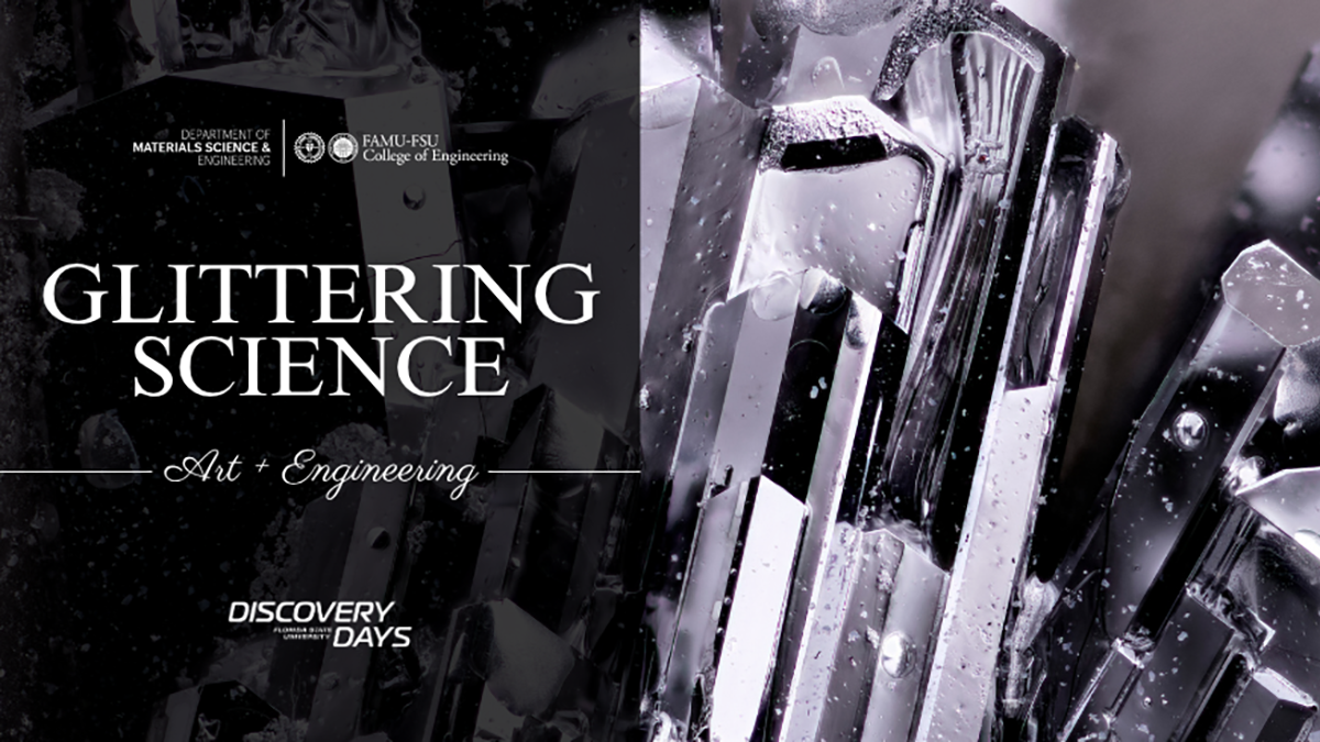
“Glittering Science” pairs crystal research with macro photography at the FAMU-FSU College of Engineering, Oct. 21, 6:30 to 8 p.m.
Assistant Professor William Meier grows crystals in his materials science and engineering lab to study fundamental physics.
College photographer Scott Holstein captures the crystals through macro photography, magnifying them up to five times their actual size.
The free event includes a lecture, reception and gallery exhibit at the IRCB.
Curiosity and creativity collide this fall as the FAMU-FSU College of Engineering invites the public to a one-of-a-kind event featuring two of its own: William Meier, assistant professor of materials science and engineering, and Scott Holstein, the college’s professional photographer.
Meier explores the secrets of physics by growing crystals in his lab, while Holstein captures their shimmering beauty through his lens, turning scientific discovery into visual art. The college is hosting a showcase of the work at the Glittering Science event on Oct. 21, 6:30 to 8 p.m., at the Interdisciplinary Research and Commercialization Building (IRCB) for a lecture, reception and gallery of work.
The Science of Crystals
“Growing these crystals is similar to making rock candy by cooling hot sugar syrup in water, but instead of water, we use molten metals like aluminum, bismuth, or zinc,” Meier said. “We mix the metals with a pinch of other chemicals in a ceramic container, heat the mixture to melt everything and then slowly cool it so crystals can grow from the liquid metal. When it works, we get shiny metal crystals.”
The payoff is fundamental science.

“We grow crystals to support scientific experiments and to answer fundamental questions about materials,” he continued. “By studying their behaviors—such as magnetism, superconductivity and how they conduct electricity—we gain insight into the underlying physics and can design new materials to solve technological challenges.”
Those crystals end up in technologies like microchips, lasers, quantum sensors and fusion reactor components. Single crystals, in particular, let researchers measure how properties such as electrical conductivity and magnetism vary with direction, revealing unique information about electron behavior within the material—something only possible with large, high-quality crystals.
Getting there isn’t always tidy.
“Some of the most beautiful crystals are those that don’t go as planned,” Meier said. “We want blocky crystals for experiments, but sometimes we get tree-shaped crystals with sparkling, thin branches that are beautiful but unusable for science.” Creating high-quality crystals takes both technical skill and creativity, he said. Reactants oxidize in air, crystals turn out too small to use, or the lab lacks a suitable molten metal for the job. “Sometimes creativity leads to a solution,” Meier said. “Other times, we move on to an easier project.”
Students are central to that process at every level. “Undergraduates grow crystals from elemental materials and run experiments to identify their properties, gaining hands-on experience with advanced equipment,” Meier said. “My graduate students typically take on more advanced synthesis projects that require careful process adjustments to obtain the desired crystals. They also travel to national laboratories to conduct advanced experiments using high-powered X-ray machines or particle accelerators.”

The crystals themselves are tiny: “0.1 to 10 millimeters, or 0.004 to 0.4 inches,” Meier said, adding that he’s always hoping for larger ones because they’re easier to work with. “Handling a crystal only three times the width of a human hair is very stressful.”
Crystal growth is a collaborative process that doesn’t end in Meier’s lab. Scientists around the world request his crystals for their own experiments, and Meier says he might ship samples to California, New York, Illinois, Tennessee or Germany for collaborators to study with specialized equipment. “Some use X-rays to analyze atomic patterns, others place crystals near nuclear reactors to examine magnetism or use lasers to study how electrons move,” Meier said. “I enjoy this aspect because I get to learn about the fascinating science they discover.”
Capturing Crystals Through the Lens
Holstein recalls the first time he saw one of Meier’s crystals under the lens. “When I saw the crystals up close, through the lens, they looked like other-worldly landscapes,” he said. “It took a macro lens that was able to magnify the crystal up to five times its actual size to see the details of this secret world.”
“Macro photography takes special gear. The lens doesn’t focus like a regular one—no focus ring, just a magnification ring. To focus, I moved the camera closer or farther from the subject. A geared tripod head helped me make tiny adjustments to frame the crystals just right. In macro photography, only a thin slice of the subject is in focus. To get a sharp image, I used focus stacking, combining several photos with different planes of focus into one image using software. To make these stacks, I used a motorized macro rail to move the camera forward in tiny, precise steps—much smaller than I could manage by hand. I used the controller to manually set the start and end points and the distance the camera should move between each shot. Once I pressed start, it used the instructions I gave it to capture the whole sequence automatically,” Holstein said.
That stacking is a necessity, not a stylistic choice, at this scale.
“At 5-to-1 magnification, just a sliver of the crystal is sharp in a shot, so stacking is a must,” Holstein said. At that magnification, his lens sat less than 2 inches from the subject—tight working quarters for a process that turned out to be more manual than expected. “I thought shooting the images would be the hardest part and that the software would handle the stacking like a breeze,” he said. “In the end, I found that the software would only get me most of the way there. The rest of the process required a lot of patience and had to be done by hand.”
Lighting the crystals took some improvising.
“I diffused a flash with a small piece of copy paper folded into a tent over the crystal,” Holstein said. “Sometimes I added a second flash with a fiber-optic cable to aim the light exactly where I needed it.” Working in such tight quarters had its own hazard: adjusting the paper tent sometimes nudged the crystal out of place, forcing him to recompose the shot. Each final image, he said, is a stack of dozens to hundreds of photos.

Glittering Science
Join Meier and his students as they showcase crystals grown over the summer, brought to life through Holstein’s macro photography, at a lecture and art exhibit.
- Oct. 21, 6:30 to 8 p.m.
- Interdisciplinary Research and Commercialization Building
- 2001 Levy Ave., Tallahassee, Fla.
The crystals featured in these photographs were made possible by support from Florida State University faculty startup funds, the FAMU-FSU College of Engineering’s EUREKA undergraduate research program, the National Science Foundation and the National High Magnetic Field Laboratory. Some materials were also developed in collaboration with researchers at the University of Tennessee and with support from the Gordon and Betty Moore Foundation. Meier’s research is further enabled by national science agencies, industry partners and the Department of Defense.
Editor’s Note: This article was edited with a custom prompt for Claude Sonnet 5, an AI assistant created by Anthropic. The AI improved clarity, structure, SEO/GEO optimization and readability, while preserving the original reporting and factual content. All information and viewpoints remain those of the author and publication. This article was edited and fact-checked by college staff before being published. This disclosure is part of our commitment to transparency in our editorial process. Last edited: 09-16-2026.
RELATED ARTICLES
Assembling polymer crystals and deciphering how they grow
Engineer One of Two Researchers Named Fellows of American Association for the Advancement of Science